The most comprehensive analysis of the 3D structure of SARS-CoV-2 to date has revealed new insight on how the virus infects human cells and replicates.
Led by Professor Sean O’Donoghue, from the Garvan Institute of Medical Research and CSIRO’s Data61, researchers compiled more than 2000 different structures involving the coronavirus’s 27 proteins. The analysis identified viral proteins that ‘mimic’ and ‘hijack’ human proteins – tactics that allow the virus to bypass cell defences and replicate.
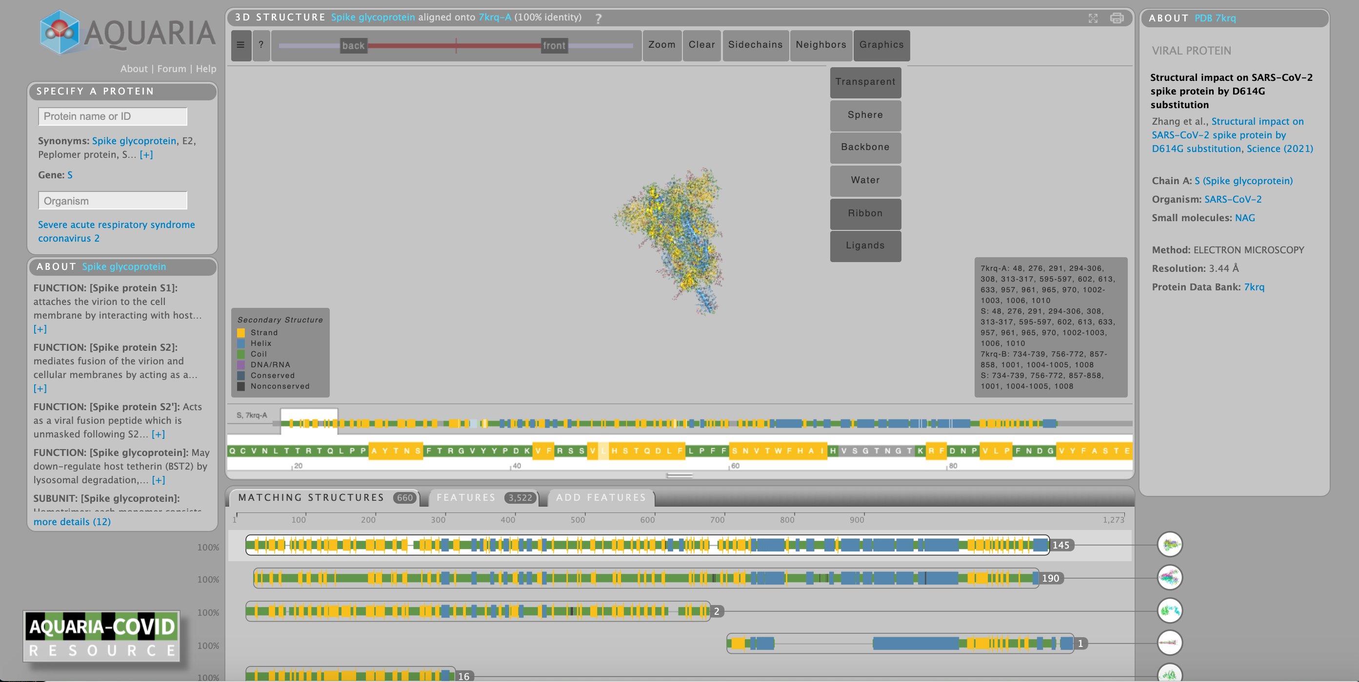
These structural models can be freely accessed from the Aquaria-COVID resource, a website designed by the team to help the research community ‘zoom in’ on potential new targets on the virus for future treatments or vaccines, and crucially investigate new virus variants.
“Our resource contains a level of detail of SARS-CoV-2’s structure that is not available anywhere else. This has given us an unprecedented insight into the virus’s activity,” says Professor O’Donoghue, first author of a paper in the journal Molecular Systems Biology detailing the team’s findings.
“Our analysis has highlighted key mechanisms used by the coronavirus; these mechanisms, in turn, may guide the development of new therapies and vaccines.”
Structural insights
To better understand biological processes, researchers determine the 3D shape of individual proteins – the building blocks that make up cells or viruses.
“3D structures of proteins provide us with atomic-resolution information on the composition of SARS-CoV-2 that is crucial for developing vaccines or treatments targeting distinct parts of the virus.
Thanks to a recent research focus on SARS-CoV-2, scientists have determined around a thousand 3D structures of the virus’s 27 individual proteins, and nearly a thousand more for related proteins,” explains Professor O’Donoghue. “However, until now there has been no easy way to bring all the pieces of data together and analyse them.”
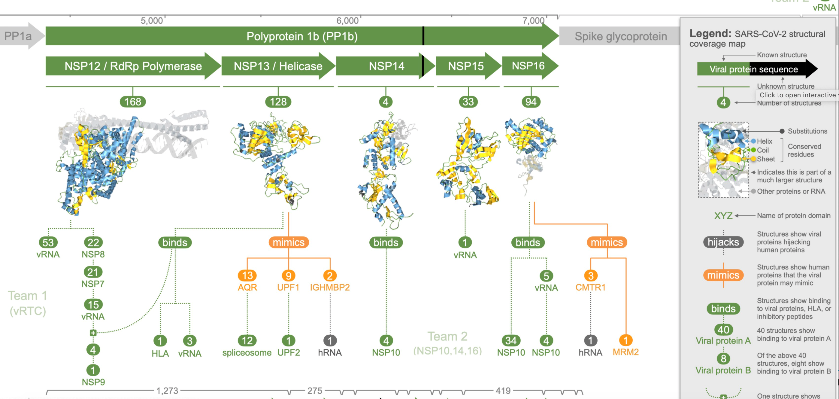
The team’s analysis revealed three coronavirus proteins (NSP3, NSP13, and NSP16) that ‘mimicked’ human proteins, which the researchers believe allows the virus to better hide from the human immune system and may contribute to the variation in COVID-19 outcomes.
The modelling also revealed five coronavirus proteins (NSP1, NSP3, spike glycoprotein, envelope protein, and ORF9b protein) that the researchers say ‘hijack’ or disrupt processes in human cells, thereby helping the virus take control, complete its life cycle and spread to other cells.
“Further, we found eight coronavirus proteins that self-assemble with each other — analysing how they assembled has provided new insights into how the virus replicates its genome. However, after accounting for overlaps, this still leaves 14 proteins that we think play key roles in infection but have no structural evidence of interaction with other viral or human proteins,” says Professor O’Donoghue.
“To make all these insights and data more accessible to researchers, we devised a new visualisation method called a structural coverage map. The map highlights what we know about SARS-CoV-2 and what is still left to uncover — it also helps scientists find and use 3D models to investigate specific research questions.”
Viral surveillance
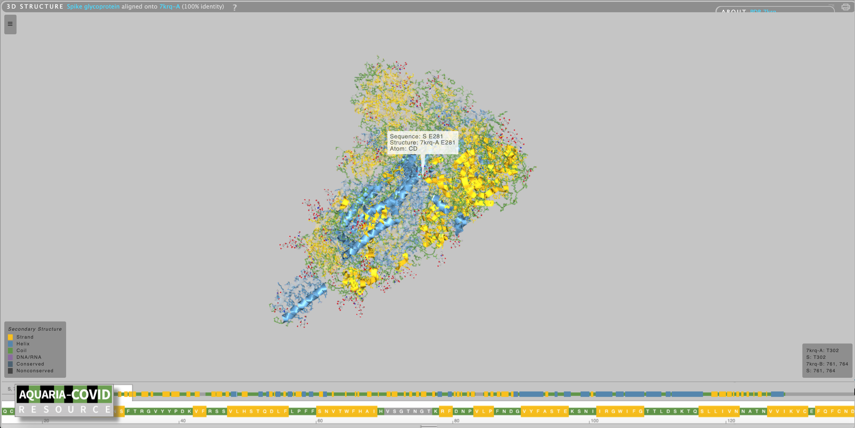
The team’s analysis reveals opportunities for further research. “Much of the coronavirus research to date has focused on the spike glycoprotein, which is the main target for current vaccines. This protein will continue to be an important target, but it’s also important we broaden our focus to other potential targets and better understand the entire viral lifecycle,” says Professor O’Donoghue.
He adds that the Aquaria-COVID resource may help researchers more easily investigate how new variants of coronavirus differ – and critically, how they may better be targeted with vaccines and treatments.
“The longer the virus circulates, the more chances it has to mutate and form new variants such as the Delta strain,” says Professor O’Donoghue. “Our resource will help researchers understand how new strains of the virus differ from each other – a piece of the puzzle that we hope will help in dealing with new variants as they emerge.”
This research was supported by the Sony Foundation Australia, Tour de Cure Australia, the Wellcome Trust, Biotechnology and Biological Sciences Research Council and the Bundesministerium für Bildung und Forschung (BMBF).
This project was a collaboration between the Garvan Institute of Medical Research, CSIRO Data61, UNSW Sydney, Weihenstephan-Triesdorf University of Applied Sciences, Technical University of Munch, The University of Dundee, and University College London.
Professor O’Donoghue is a Conjoint Professor, School of Biotechnology and Biomolecular Sciences (BABS), UNSW Sydney and a Visiting Scientist, Commonwealth Scientific and Industrial Research Organisation (CSIRO) Data61, Australia’s national science agency.
This media release was originally published here: https://www.garvan.org.au/news-events/news/3d-analysis-of-sars-cov-2-reveals-clues-on-virus-tactics